CERN70: From physics to medicine
14 November 2024 · Voir en français
Part 21 of the CERN70 feature series. Find out more: cern70.cern
Ugo Amaldi helped create a European network of cancer therapy centres using beams of ions, in particular carbon ions

Technology developed at CERN for accelerators and detectors has found many uses in areas beyond the field of particle physics, not least in medicine, where there are applications in both the diagnosis and treatment of disease.
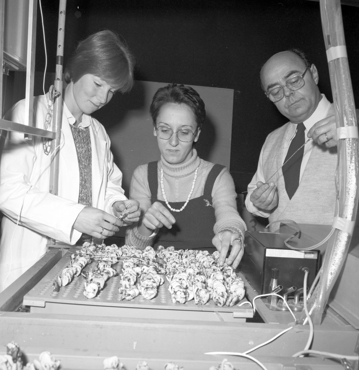
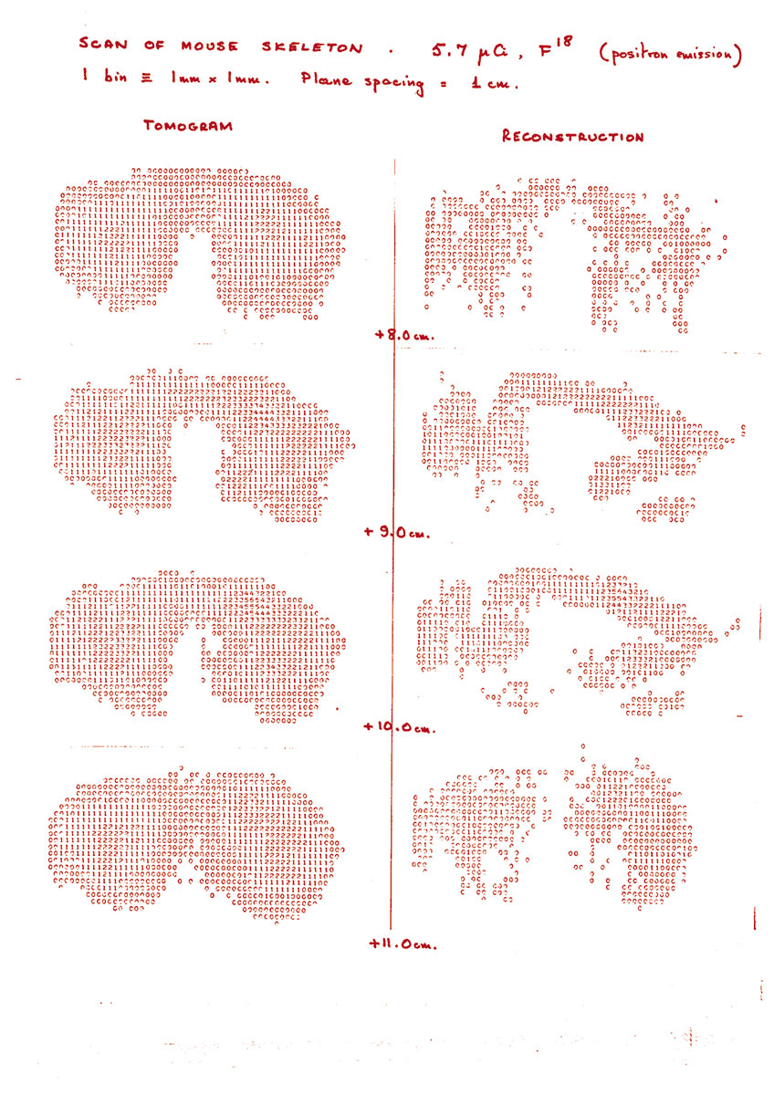
Researchers at CERN have been contributing to medical research for nearly six decades, ever since radiobiology experiments began in 1965. Three years later, Georges Charpak invented the multiwire proportional chamber, a new type of detector for which he was awarded the Nobel Prize in Physics, and which came into use for medical imaging in the mid-1970s. Improvements in detectors, electronics and materials science have enabled doctors to significantly reduce the radiation dose to the patient during the examination, while improving image quality. In an important development, a prototype “positron camera” took the first PET image of a mouse in 1977, when David Townsend developed algorithms to reconstruct data from Alan Jeavons’s detector using the radiobiology experiments of Marilena Streit-Bianchi. In 1979, David Townsend moved to the Geneva University Cantonal Hospital, where he helped build the hospital’s first positron emission tomography (PET) scanner, based on the positron camera.

Around the same time, physicists at CERN also became interested in using protons and carbon ions (atomic nuclei) for cancer therapy – an idea that dates back to 1946, when the use of protons and carbon ions was first proposed by the American accelerator physicist Robert Wilson, who later became the founder and first director of Fermilab. Protons and the nuclei of light elements, such as hydrogen and carbon, are better suited than X-rays to the treatment of some deep-seated cancers because their energy deposition (the “dose”) can be focused to cause less damage to healthy tissue.
In the late 1970s and 1980s, CERN’s accelerator experts were increasingly consulted for medical accelerator projects, in particular in the newly developing field of light ion therapy. In the 1990s, the first Medipix collaboration began and the Crystal Clear collaboration was set up to improve scintillating crystals. Both collaborations have benefited particle physics detection and medical imaging.
Then, in 1995, Ugo Amaldi, a physicist at CERN who had created the Italian TERA Foundation for radiation therapy, together with Meinhard Regler of the MedAustron project, convinced the CERN Management to start the Proton Ion Medical Machine Study (PIMMS), directed by Phil Bryant. The design of PIMMS was completed in 2000 and was later adapted by TERA to meet the needs of a centre built in Italy, CNAO (National Centre for Oncological Hadron Therapy), which began treating patients in 2011. In 2019, CERN launched the Next Ion Medical Machine Study (NIMMS) to develop cutting-edge accelerator technologies for a new generation of compact and cost-effective ion-therapy facilities.
Ongoing collaborations with medical doctors, epidemiologists and researchers are leading to new developments that help to preserve or improve our health. Examples include the role of Medipix in medical imaging, including colour X-rays, FLASH radiotherapy, the use of radioisotopes for diagnosis and treatment of diseases, machine learning and more.
Recollections
When the first patients were treated at CNAO, I felt that I’d done very interesting things in physics, but hadron therapy was the best thing I’d done in my professional life.
Ugo Amaldi

Ugo Amaldi was well known as a particle physicist at CERN when he decided to work towards the creation, in Europe, of a network of centres for cancer therapy using beams of ions, in particular carbon ions.
“My interest in medical physics dates back to the late 1950s, when I began to work at Italy’s national health institute in Rome, the Istituto Superiore di Sanità (ISS), and wrote a 700-page treaty entitled Fisica delle Radiazioni. I went to ISS instead of starting an academic career because my father Edoardo, a well-known physicist and one of CERN’s founders, had been a professor in Rome since 1937 and I wanted to distinguish myself from him.
At the ISS physics laboratory, I performed experiments in nuclear and particle physics and worked part-time in radiation physics and radiotherapy, a subject that I really enjoyed. From 1968 to 1973, I led a group of physicists from ISS working at CERN, and then – after a successful experiment at the Intersecting Storage Rings that discovered the increase with energy of the proton–proton cross-section – in 1973, I was offered a staff position at CERN. One thing led to another and, by 1981, I had initiated and become the spokesperson for DELPHI, one of the four LEP experiments.
By 1991, following the first results and a much-quoted paper on the supersymmetric unification of the fundamental forces, DELPHI was running very well. But it wasn’t obvious to me what I should do next. I was 57 years old and I knew I wouldn’t be an active experimental physicist when the LHC came online. Still, I wanted to do something new and directly useful at the frontier of what could be done with particle accelerators. I therefore decided to go back to radiation physics; a few years later, my wife Clelia said, “You went back to your first love”.
In fact, the idea of using physics to help cure people still appealed to me so, in 1991, I wrote, with Giampiero Tosi, a well-known Italian medical physicist, a proposal for a cancer therapy centre based on beams of carbon ions (and of protons), a treatment modality that, shortly after, I called “hadron therapy”. One year later, we created the TERA Foundation to raise funds to cover personnel costs for the design of this centre, for which we received help from the Italian Institute for Nuclear Physics (INFN).

Then, as now, the main tool for cancer therapy was radiotherapy with X-rays, but dedicated proton centres were also being built. However, I was interested in some results obtained at the Lawrence Berkeley Laboratory and in papers written by Gerhard Kraft of GSI. About 1% of tumours treated with X-rays turn out to be resistant to both X-rays and protons, but it appeared that these stubborn tumours might respond if they were bombarded with the nuclei of carbon atoms. Each carbon nucleus leaves 20 times more energy than a proton in a cell and thus produces multiple, irreparable breaks of the DNA double helix. For this reason, carbon beams are effective in controlling “radioresistant tumours”, such as soft-tissue sarcomas, bone sarcomas and some cancers of the brain, lung and salivary glands.
At the end of 1995, with Meinhard Regler of MedAustron, we drew the interest of the CERN Management to the design of an ion synchrotron optimised for such a medical application. This became PIMMS, the Proton Ion Medical Machine Study, on which physicists and engineers from CERN, TERA and the MedAustron project worked for four years under the direction of Phil Bryant. In parallel, with TERA, I proposed that the Italian government finance a treatment centre based on an improved version of the PIMMS synchrotron, later called the PIMMS/TERA project. As the centre would cost more than 100 million euros, this was a hard sell, but the Italian government finally put up the money in 2001. In 2003, the CNAO Foundation, with its team of 26 physicists and engineers took over the project. Sandro Rossi, who had been the TERA Technical Director for almost ten years, became the CNAO Technical Director and, later, its Director-General. With the help of INFN, in autumn 2005, the construction of the Centre started in Pavia, an ancient university town close to Milan. In 2011, the first proton irradiation took place and, in 2012, exactly 20 years after the creation of TERA Foundation, the first patient was treated with a beam of carbon ions.
When the first patients were treated at CNAO, I felt that I’d done very interesting things in physics, but hadron therapy was the best thing I’d done in my professional life.
I am now President Emeritus of CNAO, and I have the satisfaction of seeing that the Centre is still growing. More than 5000 patients have been treated, 55% with carbon ions, and more and more patients are children: in 2023, 62 paediatric tumours were irradiated. Moreover, a proton gantry is being installed and a novel boron neutron capture therapy system will be operative in 2026. Overall, one can say that CNAO, as a spin-off of CERN and TERA, fully justifies my motto “Physics is beautiful and useful”.
This video shows how technologies developed for particle accelerators, detectors and analysis have found their way into medical applications.
---
This interview is adapted from the 2004 book “Infinitely CERN”, published to celebrate CERN’s 50th anniversary, and was updated with the help of Ugo Amaldi in 2024. Ugo was one of the speakers at the recent CERN70 public event “From particle physics to medicine”. A new PET digital learning module is available now for high-school students to help save a virtual patient, find out more via cern.ch/petlearningmodule.
